+91-98777-81556 info@sepalhealthcare.com

Elbow 2 Column System 2.7/3.0 mm

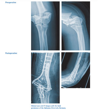
Elbow Plates 2.7/3.0 comprise bone plates and screws for treatment of fractures at the distal Humerus and the proximal ulna, in addition to the necessary accessories for internal fixation. The implants contain specialised features that result in quicker healing and early mobilization of the patient.

Elbow 2 Column Plates 2.7 and 3.0 mm systems are used for the temporary fixation, correction or stabilization of the distal humerus as well as the olecranon. The implants are intended for single use in the patients. The system contains olecranon plates, coronoid plates, posterolateral distal humerus plates, lateral column plates, medial column plates and the supporting screws. 

The main advantages of the Plate include:

Following are some of the advantages of the Elbow 2 Column System -

Surgical Technique

Pre-surgical -

The surgeon goes through the investigations of the patient, such as X-Ray, MRI and CT/CAT, and counsels the patient and the attendees regarding the procedure and the expected outcomes of the surgery.

Distal Humerus Plates 2.7/3.0mm

These Distal Humerus Plates are pre-contoured offering anatomical compatibility, and thus can be used as a template for restoring fracture fragments.
Below is the surgical technique used in case of Distal Medial and Distal Dorsolateral Humerus and Distal Lateral Humerus Plates:

Preparing the Plate:

The drill guide has to be inserted in the most distal hole of the medial plate. It has to be positioned in a way that the opposite pointer is lateral to the desired point of exit of the next screw.

Insertion of Cortical Screws & Locking Screws:

Using the double drill guide, a cortical screw 3.5 mm is inserted in the oblong hole. The length of the screw is determined using a depth gauge. Using a screwdriver or low power drill, locking screws are inserted until optimum locking is achieved. Locking compression screws of appropriate length also need to be inserted using the same technique, with suitable equipment.

Prophylactic Antibiotics –

To treat the prevailing infections and to avoid the postoperative infections, the doctor prescribes antibiotics prior to the surgery. Administering antibiotics also ensures better healing post the surgery.

Positioning the Patient:

The patient is positioned in a lateral or prone position, with the affected arm supported over bolsters. A longitudinal incision is usually done through the posterior side, passing the olecranon on the radial side. It is necessary to protect the ulnar nerve.

Reduction of Fracture and Preliminary Fixation:

The fracture is reduced and temporarily fixed using K-wires or independent lag screws. The medial plate is inserted and positioned. The plate is fixed to the bone using K-wires. When a cortical screw is used in the oblong hole for primary fixation, it enables required corrections in plate positioning. Fluoroscopy can be used to confirm anatomic reduction and plate positioning.

Olecranon Plate 2.7 / 3.0 mm

Positioning the Patient:

The patient is positioned in lateral or prone, with the arm supported over bolsters or tourniquet. Usually, the posterior access lateral to the elbow is preferred. Incision is made about 5cm distal to the supracondylar area. Care should be taken to avoid damaging the ulnar nerve.

Preparing the Plate:

Using an aiming device, a drill guide is inserted through the most proximal hole. Intra-operative bending of the plates is not necessary as these are anatomically pre-contoured. Only in case of necessity, the plates may be contoured using bending irons.

Reduction and Preliminary Fixation:

The fracture is reduced and fixed preliminarily using K-wires and lag screws. The anatomic reduction can be confirmed using fluoroscopy. It has to be taken care that no screws are penetrating the articular surfaces.

Insertion of Locking Screws and Final Fixation of Fracture:

The most distal metaphyseal plate hole is secured with a screw. Cortical and locking screws of appropriate measurement are used to perform fixation of the plate shaft. Confirmation regarding the final positioning of the screws is done through fluoroscopy, and screw length and position are altered, if necessary.

X
Open chat
Hello , How Can We Help You ?