+91-98777-81556 info@sepalhealthcare.com

Clavicle Hook Plate – 3.5 mm

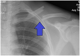
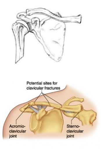
Simply put, the clavicle is the collar bone. The clavicle is situated between the ribcage (sternum) and the shoulder blade (scapula). The clavicle attaches our upper limb to the body.


medical articular clavicle fracture

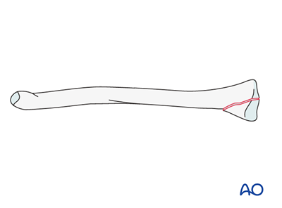
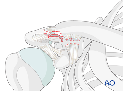
Partial articular fractures of the medial end of the clavicle are classified as AO/OTA 15.1B fractures.
Definition: The medial end is the segment of the clavicle involving the costoclavicular ligament.

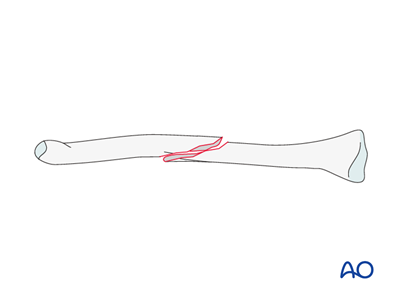
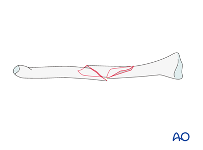
Diaphyseal simple, spiral clavicle fracture

The AO/OTA classification does not further subdivide simple fractures of the diaphysis into subtypes (spiral, oblique, or transverse), but the treatment may vary depending of the fracture configuration..
They are all classified as AO/OTA 15.2A fractures.
Definition: The diaphysis of the clavicle extends from the attachment of the coracoclavicular ligament laterally to the costoclavicular ligament medially.

Diaphyseal simple, transverse clavicle fracture

The AO/OTA classification does not further subdivide wedge fractures of the diaphysis into subtypes (spiral, bending or fragmented), but the treatment may vary depending of the fracture configuration.
They are all classified as AO/OTA 15.2B fractures.
Definition: he diaphysis of the clavicle extends from the attachment of the coracoclavicular ligament laterally to the costoclavicular ligament medially.

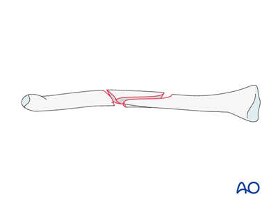
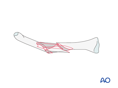
Diaphyseal wedge, fragmentary wedge clavicle fracture

The AO/OTA classification does not further subdivide wedge fractures of the diaphysis into subtypes (spiral, bending or fragmented), but the treatment may vary depending of the fracture configuration.
They are all classified as AO/OTA 15.2B fractures.
Definition: The diaphysis of the clavicle extends from the attachment of the coracoclavicular ligament laterally to the costoclavicular ligament medially.

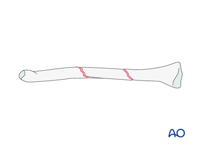
Diaphyseal multifragmentary, intact segmental clavicle fracture

The AO/OTA classification does not further subdivide multifragmentary fractures of the diaphysis into subtypes (fragmented spiral, intact segmental, fragmented segmental), but the treatment may vary depending of the fracture configuration.
They are all classified as AO/OTA 15.2C fractures.
Definition: The diaphysis of the clavicle extends from the attachment of the coracoclavicular ligament laterally to the costoclavicular ligament medially.

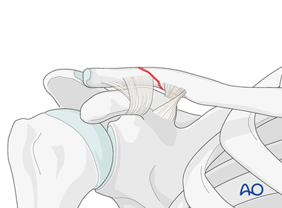
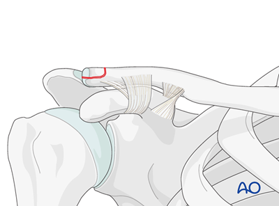
Lateral, undisplaced fracture with CC intact, extraarticular clavicle fracture

An impacted or undisplaced extra articular fracture with intact coraco-clavicular ligaments is classified as AO/OTA 15.3A(a) fracture. The ligaments may be minimally stretched.
Definition: Distal fractures of the clavicle involve the clavicle lateral to a perpendicular line to the medial cortex of the coracoid base.
These fractures typically occur from a direct blow to the point of the shoulder such as from a fall in which the patient lands directly on the shoulder.
Epidemiology of these fractures is bimodal with young active patients sustaining injuries from sporting events, bicycling accidents, and motor vehicle collisions. Older patients typically sustain these injuries in falls from a standing height.
The history should include the age, activity level, comorbidities, and mechanism of injury as these will be used in the decision making regarding surgery. A thorough examination of the involved limb (and patient, if there is a high energy mechanism present) is mandantory prior to embarking on treatment.
Radiographic evaluation of a distal clavicle fracture includes an anterior posterior radiograph centered on the distal clavicle and a "upshot" or Zanca view which is angled 20 degrees cephalad: this allows the profile of the fracture to be seen clearly free of the scapula and rib cage. CT imaging is often obtained to assess better the fracture in the axial plane.

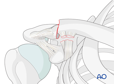
Lateral, displaced fracture with CC disrupted, articular clavicle fracture

A fracture which starts medial to the CC ligaments and runs superio laterally into the AC joint is classified as an AO/OTA 15.3B(c) fracture.
Definition : Distal fractures of the clavicle involve the clavicle lateral to a perpendicular line to the medial cortex of the coracoid base.
Displaced fractures demonstrate swelling, bruising, and a deformity with apparent superior migration of the shaft fragment as the shoulder sags downwards and medially.

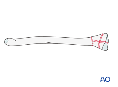
Medial, complete articular clavicle fracture

Complete articular fractures of the medial end of the clavicle are classified as AO/OTA 15.1C fractures.
Definition: The medial end is the segment of the clavicle involving the costoclavicular ligament.

Diaphyseal simple, oblique clavicle fracture

The AO/OTA classification does not further subdivide simple fractures of the diaphysis into subtypes (spiral, oblique, or transverse), but the treatment may vary depending of the fracture configuration. They are all classified as AO/OTA 15.2A fractures.
Definition: The diaphysis of the clavicle extends from the attachment of the coracoclavicular ligament laterally to the costoclavicular ligament medially.

Diaphyseal wedge, intact spiral wedge clavicle fracture

The AO/OTA classification does not further subdivide simple fractures of the diaphysis into subtypes (spiral, oblique, or transverse), but the treatment may vary depending of the fracture configuration.
They are all classified as AO/OTA 15.2A fractures..
Definition: The diaphysis of the clavicle extends from the attachment of the coracoclavicular ligament laterally to the costoclavicular ligament medially.

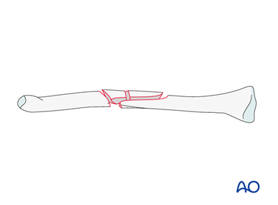
Diaphyseal multifragmentary, fragmentary spiral clavicle fracture

The AO/OTA classification does not further subdivide multifragmentary fractures of the diaphysis into subtypes (fragmented spiral, intact segmental, fragmented segmental), but the treatment may vary depending of the fracture configuration.
They are all classified as AO/OTA 15.2C fractures.
Definition: The diaphysis of the clavicle extends from the attachment of the coracoclavicular ligament laterally to the costoclavicular ligament medially.

Diaphyseal multifragmentary, fragmentary spiral clavicle fracture

The AO/OTA classification does not further subdivide multifragmentary fractures of the diaphysis into subtypes (fragmented spiral, intact segmental, fragmented segmental), but the treatment may vary depending of the fracture configuration.
They are all classified as AO/OTA 15.2C fractures.
Definition: The diaphysis of the clavicle extends from the attachment of the coracoclavicular ligament laterally to the costoclavicular ligament medially.

Lateral, undisplaced fracture with CC intact, articular clavicle fracture

An impacted or undisplaced articular fracture with intact coraco-clavicular ligaments is either classified as AO/OTA 15.3B(a) in case of a partial articular fracture, and AO/OTA 15.3C(a) in case of a complete articular fracture. The ligaments may be minimally stretched. These fractures are rare and typically stable.
Definition: Distal fractures of the clavicle involve the clavicle lateral to a perpendicular line to the medial cortex of the coracoid base.
These fractures typically occur from a direct blow to the point of the shoulder such as from a fall in which the patient lands directly on the shoulder.
Epidemiology of these fractures is bimodal with young active patients sustaining injuries from sporting events, bicycling accidents, and motor vehicle collisions. Older patients typically sustain these injuries in falls from a standing height.
The history should include the age, activity level, comorbidities, and mechanism of injury as these will be used in the decision making regarding surgery. A thorough examination of the involved limb (and patient, if there is a high energy mechanism present) is mandantory prior to embarking on treatment.
Radiographic evaluation of a distal clavicle fracture includes an anterior posterior radiograph centered on the distal clavicle and a "upshot" or Zanca view which is angled 20 degrees cephalad: this allows the profile of the fracture to be seen clearly free of the scapula and rib cage. CT imaging is often obtained to assess better the fracture in the axial plane.

Lateral, displaced fracture with CC disrupted, extraarticular clavicle fracture

A fracture which starts lateral to the CC ligaments and runs superiorly and does not affect the AC joint surface is classified as an AO/OTA 15.3A(c) fracture.
Definition :Distal fractures of the clavicle involve the clavicle lateral to a perpendicular line to the medial cortex of the coracoid base.
Displaced fractures demonstrate swelling, bruising, and a deformity with apparent superior migration of the shaft fragment as the shoulder sags downwards and medially.

Treatment:

Treatment of clavicle fracture depends on the severity of the fracture. If the bones that are fractured have not dislocated and are still aligned correctly, the fracture may not need a surgical intervention.

The non-surgical approach follows the below mentioned ways of treatment –

Treatment using Clavicle Hook Plate:

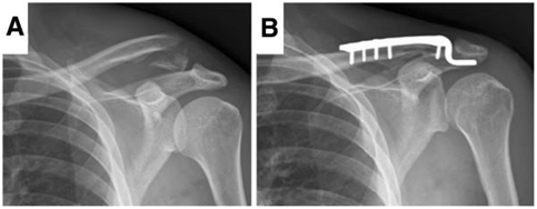
The primary use of a Clavicle Hook Plate is fixation of lateral (distal) clavicle fractures and acromioclavicular joint injuries such as dislocations. This system of plate and screws facilitates early rotational mobility of the shoulder. These plates prove to be effective fixative implants for unstable fractures of the distal clavicle.

The salient features of Clavicle Shaft Plate include:

These plates are an excellent tool for fracture reduction and fixation, in order to restore the normal anatomical functioning. Following are some of the advantages of the Clavicle Hook Plate –


Positioning the Patient and Approach:

The patient is usually positioned on a beach chair position on a radiolucent operating table. The head of the patient needs to be turned away from the side which is being operated. A sandbag is kept under the thoracic spine to enable the scapula to fall backward, enabling easier repositioning and reduction of the fracture.
In general, a superior or trans-acromial incision is made which exposes the delto-trapezial fascia. Care needs to be taken not to injure the supraclavicular nerves and arteries.

Temporary Fixation of the Plate:

The implant is positioned using a K-wire, after its position has been verified using an image intensifier. The K-wire is drilled through the drill sleeve in the distal hole to fix the plate. By aligning the medial and lateral fracture fragments with the implant using forceps, the final fixation can be carried out.

Insertion of Screws:

Based on the type and degree of fracture, appropriate screw or combination of screws will be selected. Usually, a combination of locking and cortex screws is utilised. Using a 2.5 mm drill bit, a 3.5 mm drill guide is inserted into the threaded hole and to drill for the gliding hole. Also, the appropriate length of the cortex screw is determined using the depth gauge.
Using a 2.8 mm drill bit, the screw is pre-drilled through both the cortices and the locking screw is also inserted. The procedure is repeated until all the preset holes are used, and final checking is done manually by tightening all the screws.

Reduction of Fracture and Temporary Fixation:

With the help of a K-wire or pointed reduction forceps, the fracture is reduced temporarily.

Determining the Plate Length Hook Size:

Due to the availability of the sizing templates, appropriate size of the hook is determined. After determining the size, the appropriate implant is selected. After placing the plate shaft on the clavicle, the hook’s end should be touching the underside of the acromion. The correct anatomic alignment of the clavicle and the acromion has to be restored. Using a C-arm, the full arm rotation is verified.The position of the plate can be verified using AP and axial C-arm.
There might be situations where the plate might have to be contoured to match the patient’s anatomy. This can be done usinga suitable 2.7 mm bending iron or 2.7 mm bending pliers. Also, it is imperative that the sizing templates should not be bent or contoured.

X
Open chat
Hello , How Can We Help You ?